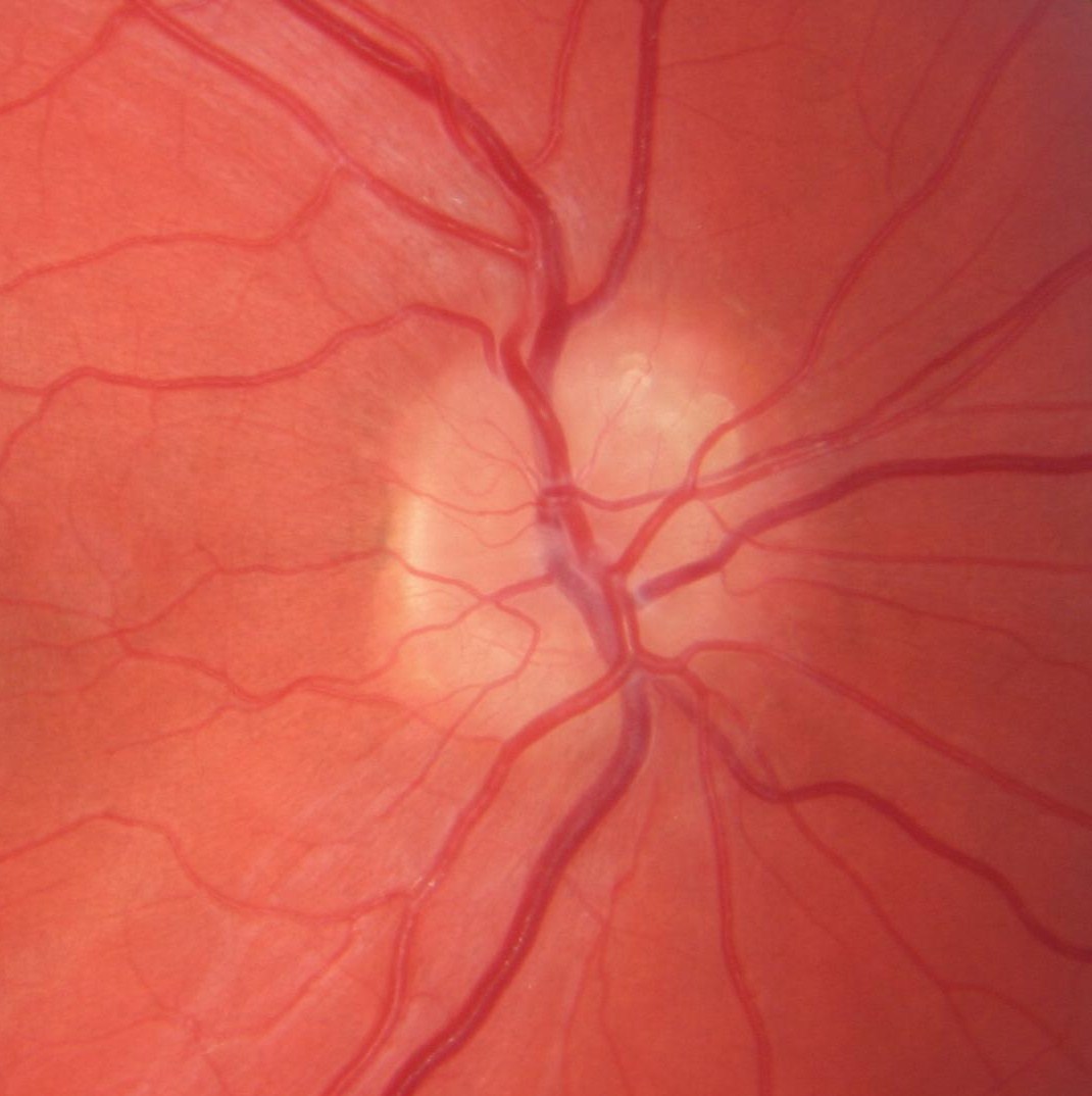
Optic Disc Anatomy. Web the optic disc is an elevation on the medial aspect of the retina where the sensory fibers and retinal vessels pass. Web optic disc anatomy. There are two main areas in the retina: The macula and the peripheral retina. Web learn about the anatomy and function of the optic disc, the entrance of the optic nerve and the blind spot in the visual field. There is a layer of tissue at the back of each eye, opposite to the pupil, called the retina. The optic disc is devoid of. Web the optic nerve head (also known as the optic disc) is approximately 1.5 mm wide and is also associated with a. This tissue is responsible for taking the light that enters the eye and turning it into the images you see. Web in the adult, the axons of about 1.2 million retinal ganglion cells converge at the optic disc to form the optic nerve. Web the structure around the optic nerve where it enters the back of the eye. Read an overview of general eye anatomy to learn how the parts of the.
from
The optic disc is devoid of. Web optic disc anatomy. Web the optic disc is an elevation on the medial aspect of the retina where the sensory fibers and retinal vessels pass. There is a layer of tissue at the back of each eye, opposite to the pupil, called the retina. This tissue is responsible for taking the light that enters the eye and turning it into the images you see. Web learn about the anatomy and function of the optic disc, the entrance of the optic nerve and the blind spot in the visual field. Web in the adult, the axons of about 1.2 million retinal ganglion cells converge at the optic disc to form the optic nerve. Web the optic nerve head (also known as the optic disc) is approximately 1.5 mm wide and is also associated with a. Read an overview of general eye anatomy to learn how the parts of the. There are two main areas in the retina:
Optic Disc Anatomy Web learn about the anatomy and function of the optic disc, the entrance of the optic nerve and the blind spot in the visual field. Web optic disc anatomy. Web learn about the anatomy and function of the optic disc, the entrance of the optic nerve and the blind spot in the visual field. There is a layer of tissue at the back of each eye, opposite to the pupil, called the retina. Read an overview of general eye anatomy to learn how the parts of the. This tissue is responsible for taking the light that enters the eye and turning it into the images you see. Web in the adult, the axons of about 1.2 million retinal ganglion cells converge at the optic disc to form the optic nerve. Web the optic disc is an elevation on the medial aspect of the retina where the sensory fibers and retinal vessels pass. There are two main areas in the retina: Web the structure around the optic nerve where it enters the back of the eye. The macula and the peripheral retina. The optic disc is devoid of. Web the optic nerve head (also known as the optic disc) is approximately 1.5 mm wide and is also associated with a.